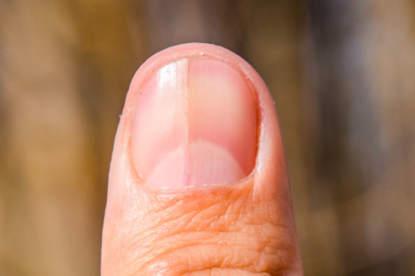
In the matrix, fingernail is produced at a rate of 0.1 mm per day. Swelling in paronychia or vascular changes in collagen vascular disease can be more easily appreciated in the nail fold where the dermis is thinned.

Each change can produce a characteristic pattern of disruption. When examining each component of the nail system, realize that the delicate interaction between vascular, melanocytic, and regenerative structures can easily be disturbed by a change in the body's normal homeostasis. Step 5: Return to the nail plate and examine it for abnormalities in color and shape. Step 4: Examine the hyponychium for abnormalities in color and shape. Step 3: Examine the nail bed for abnormalities in color and shape. Step 2 Examine the lunula for abnormalities in color and shape. Step 1: Examine the nail folds for abnormalities in color and shape. Each component of the nail is examined sequentially, pausing at each location for the detection of abnormalities in color or shape: For the nail, the digit is held in the observer's hand so that the nail is closest for observation.


A systematic examination of the nail is equally imperative. At each location, inspection, palpation, and auscultation are performed. For the heart, there is a proper position for examination, at the patient's right side, and a proper sequence in examination, beginning at the upper right sternal border and sequentially moving to the apex. Examination of the nail in a systematic manner is not unlike the systematic examination of the cardiovascular system.


 0 kommentar(er)
